Avascular Necrosis (AVN) of the Hip (also known as osteonecrosis) occurs when the blood supply to the bone of the femoral head (the ball part of the hip joint) is disrupted. This typically leads to death of the bone cells (osteocytes) in a localized area of the top of the femoral head/ball leading to collapse of the affected bone and its associated joint surface. This irreversible damage generally leads to a progressive arthritis (often quite rapid) of the hip with pain, stiffness and loss of function for walking, bending etc.
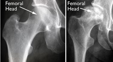
Figure 1. Normal femoral head (left) and bone changes in an avascular femoral head (right).
Avascular necrosis is a much less common condition affecting the hip than either osteoarthritis or inflammatory arthritis. In saying this AVN can affect the bone around other joints such as the knee and shoulder, but the hip is the most common joint with this condition.
In most cases, the cause of this loss of blood supply is unknown or termed “idiopathic”. There are however a number of risk factors that make it more likely for someone to develop this condition:
- Traumatic Injury – Hip dislocations and certain types of hip fracture can damage the blood supply to the femoral head.
- Alcohol Intake – Excessive alcohol consumption over a long period of time can be a causative factor. It is not known how this occurs.
- Corticosteroid Medication – Cortisone/Prednisolone medications for treatment of airways disease or inflammatory arthritis/bowel disease or other conditions. Long term use carries a higher risk of developing AVN but even short term relatively high dose administration has been associated with this condition. Again it is not known how these drugs lead to AVN. Chemotherapy agents (used to treat various cancers) can lead to the development of AVN in a similar manner.
- Other Medical Conditions – Blood disorders like Thalassaemia and Sickle Cell Disease and Metabolic conditions like Gauchers disease can cause small clots to form in the tiny arteries supplying blood to the femoral head thereby blocking blood flow to part of the bone. An unusual condition called Caisson’s Disease (seen in deep sea divers experiencing “the bends”) causes the release of nitrogen bubbles into the arterial circulation which can block the blood vessels of the femoral head in a similar manner. There are many other conditions which can be associated with AVN of the Hip but they are quite rare.
Avascular necrosis of the hip usually occurs in adults in the range from 50-70 years but can occur at any age, even in children occasionally. AVN of the hip develops in stages and early on there are often no symptoms with the condition only being picked up incidentally on a scan or other radiological investigation. As the condition progresses and there are bone changes, most patients will experience pain which is usually felt in the groin area but may radiate into the thigh or very occasionally into the buttock. Once the bone of the femoral head starts to collapse, the pain usually starts becoming more severe, affecting all weight bearing activities but also occurring with leg movements and bending. With end stage AVN, the hip is often painful at rest, movement is restricted and activity is severely limited. AVN can progress through these stages quite rapidly over a period of just a few months or it may take 12 – 18 months. This is in contrast to osteoarthritis of the hip which is a generally slowly progressive condition that takes years to develop.
Figure 2. The Progressive Stages of Avascular Necrosis of the Femoral Head.
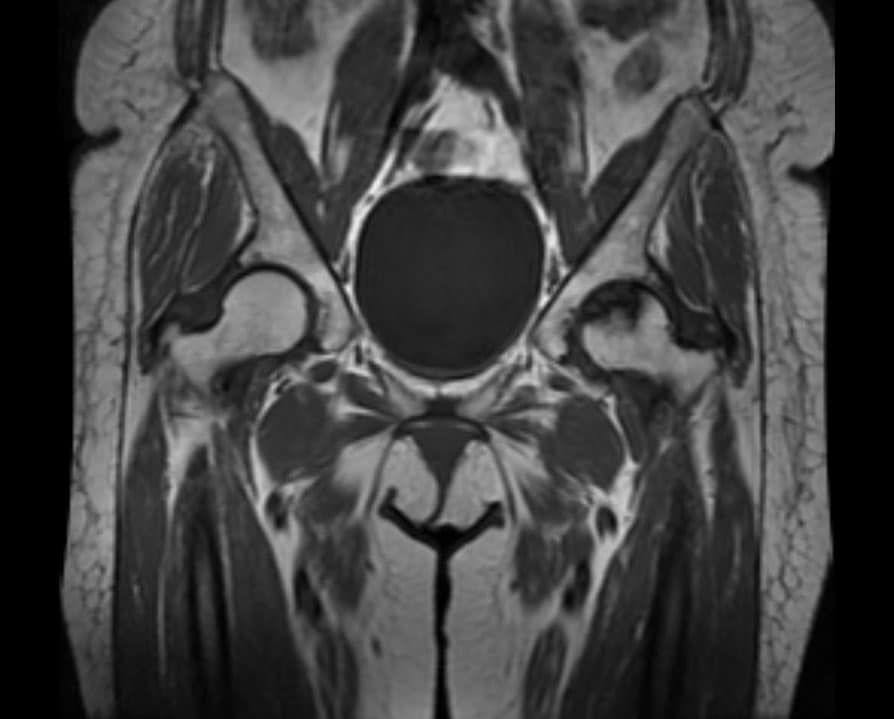
The diagnosis of Avascular necrosis of the hip is usually made after obtaining xrays or scans of the hip but sometimes can be quite difficult. The diagnosis may be suspected in a patient with a painful hip with restricted movement who may have one of the recognized conditions or risk factors (outlined above) sometimes associated with AVN. Plain xrays of the hip will usually show bone changes (increased density or whiteness of the bone in a localized segment of the femoral head, early collapse – a crescent sign or more advanced collapse) once the condition is established. Nuclear bone scans will sometimes pick up this condition but are often non specific. MRI scan is probably the gold standard investigation for this condition if it is suspected but plain xrays of the hip are normal. MRI has the capability to pick up very early stage AVN before symptoms have started as well as more advance disease and is useful for determining how much bone is involved by the condition. It is also extremely useful for evaluating the opposite hip for this condition as AVN can develop in both hips, particularly in those individuals where a systemic or general risk factor is present, for example alcoholism, steroid use, blood conditions etc.
Figure 3. An MRI scan of the hips showing a normal femoral head (left) and typical bone changes of AVN (femoral head on right side of the scan).
The treatment of avascular necrosis depends on a number of factors – individual factors, how much bone is affected and most importantly the stage of the condition. Whilst non operative or conservative measures may be appropriate for some patients with AVN, most will require some form of surgical intervention. Painkilling/analgesic and anti-inflammatory medications and the use of crutches can reduce the symptoms and possibly slow down the progression of AVN, but rarely results in resolution. Sometimes these measures are used when the diagnosis is not clear – it can sometimes be difficult to differentiate AVN of the hip from transient idiopathic osteoporosis (TIO) or stress/insufficiency fractures of the femoral head. Follow up with serial MRI scans is usually required to ascertain the correct diagnosis.
Surgical treatment involves either a procedure called Core Decompression (also known as a “forage” procedure) or Total Hip Replacement.
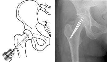
Core Decompression is a procedure that involves the drilling of one large or several small holes into the affected area of the femoral head to relieve pressure within the bone and to create channels for new blood vessels to grow into the “avascular” bone in an attempt to re-establish a new blood supply. This procedure is only relevant when AVN is diagnosed in its very early stages – often before symptoms and certainly before changes are seen on plain xray. Unfortunately this procedure is not successful in all cases. Sometimes bone grafting is undertaken in addition to decompressive surgery to try and lessen the chances of bone collapse but so far this has not conclusively been shown to improve outcomes in AVN.
Figure 4. Core Decompression for Avascular Necrosis of the Femoral Head
Unfortunately for the majority of patients diagnosed with AVN of the hip, Total Hip Replacement (THR) remains the only treatment that is able to satisfactorily deal with the damaged bone and joint surface of the femoral head. Similar to end stage osteoarthritis, it involves replacing the ball and the socket of the joint with artificial implants which have bearing surfaces which provide for generally very satisfactory pain relief, restoration of movement and significant improvements in function (walking, bending and activities of daily living/simple recreations etc.). THR is regarded as one of the most successful operations in all of medicine.
Source: https://www.parkclinic.com.au/avascular-necrosis-of-the-femoral-head/


