BLOG

Medications and Surgery

medication1

What medications need to be ceased before a surgical procedure?

This is a very common question that crops up before surgery. Medications that increase the possibility of complications such as bleeding or infection or thrombosis ideally need to be ceased prior to undertaking surgical procedures. Frequently these medications are taken for important medical conditions and as such, at times, consultation between your treating surgeon and the prescriber of the medication (GP or physician) will need to take place. This is to devise a plan for how and when the medication should be ceased and whether any covering medication is required and importantly when medication needs to be recommenced after surgery and how this will take place.

Blood Thinning Agents

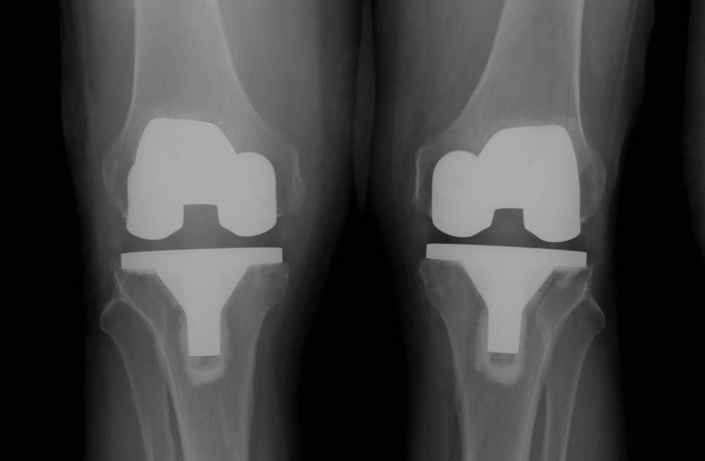
The issue with these drugs is their ability to increase surgical blood loss. This is clearly important in major open cases such as joint replacement surgery, where blood loss can be significant and ongoing loss after surgery can occur if normal clotting mechanisms are impaired. Bleeding can even be a problem in minimally invasive surgery such as arthroscopy. Usually the joint is filled with a clear fluid (normal saline) which provides clear and easy vision to allow the arthroscopic procedure to take place. Bleeding inside the joint (and particularly bleeding that is difficult to control) creates a “tomato soup” effect rather than a clear visual field. If this is bad and can’t be cleared, the procedure may need to be abandoned.

A. Aspirin and Anti-Inflammatory Drugs

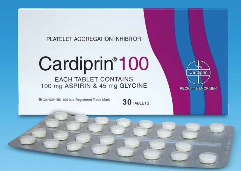
Common medications leading to blood thinning include aspirin and non steroidal anti-inflammatory drugs (NSAID). Examples are Astrix, Cardiprin (aspirin) and Voltaren, Naprosyn, Nurofen, Mobic and Celebrex (NSAID). These affect platelets and their function for about a week. It is recommended that these agents are stopped 7 – 10 days before surgery.

cardiprin-100-tab6002pps0

B. Platelet Aggregation Inhibitors

More powerful anti-clotting agents like clopidogrel affect platelets also but their effect is longer lasting than for aspirin/NSAIDs and it is recommended that these agents are stopped at least 14 days prior to a surgical procedure. Examples include Plavix and Iscover.

These agents are often prescribed in patients with coronary artery stents in the first 12 months after insertion to prevent stent blockage from occurring. In this time period it is probably better to avoid elective surgery or if surgery needs to be undertaken, advice is sought from the treating cardiologist.
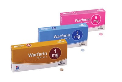
C. Warfarin

Drugs like Warfarin (Coumadin) which effect clotting pathways are usually stopped 5 – 7 days before surgery as this is the sort of time required for clotting to normalize. This can be checked with a simple blood test. Warfarin is often given to prevent stroke in individuals with cardiac arrhythmias such as atrial fibrillation, as a treatment for venous thrombosis and in patients with certain types of artificial cardiac valves. Sometimes in these circumstances Warfarin or similar agents need to be replaced with agents that have a short duration of action (Clexane/Heparin 24 hours) to provide anticoagulation cover until the day before surgery. If the matter is urgent Warfarin can be reversed quickly with VitaminK or infusion of fresh frozen plasma.

1-warfarin

D. Oral Heparin-like Agents

Newer agents such as Rivaroxaban and Epixaban are becoming more frequently used instead of Warfarin because of their relatively short duration of effect and the fact that they don’t need to be constantly monitored with regular blood tests. These agents can generally be safely ceased the day before surgery.

E. Glucosamine/Chondroitin

It has been suggested in some studies that Glucosamine and Chondroitin and large doses of Omega-3 oils (fish oil/krill oil) can adversely affect clotting and should be ceased 14 days before surgery.

I’m generally happy for all these agents to be recommenced a couple of days after surgery, provided that ongoing blood loss has ceased and the patient’s condition is medically stable.


Other Medications Affecting Clotting

The oral contraceptive pill (OCP) and hormone replacement therapy (HRT) have been shown to increase clotting and therefore the risk of developing deep vein thrombosis. These agents should be ceased 1 week prior to any surgery where there is a significant risk of deep vein thrombosis – total knee and hip replacement procedures or major pelvic surgery.


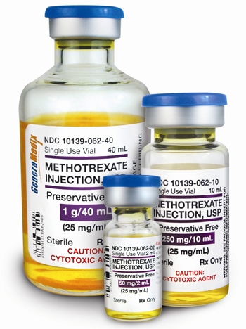
Immunosuppressive Medications

These medications include Methotrexate, Humira, Arava etc and even Prednisolone or Cortisone by mouth and are often prescribed in individuals with auto-immune disorders such as rheumatoid, psoriatic and other inflammatory arthritis conditions. In a surgical setting, these drugs can make the body more susceptible to developing infection and impair the ability to fight infection. These drugs also retard normal skin and soft tissue healing. As such they pose a hazard, particularly in patients undergoing major surgery such as joint replacement procedures. These drugs should be ceased at least one half-life before surgery (usually 1-2 weeks) and should not be recommenced until wound healing has taken place (usually after post operative review at 2 weeks).

bioniche_Methotrexate


Drugs Affecting General Health

Cigarette smoking has been shown to retard bone healing. Smoking also increases anaesthetic risk and risk of developing post operative chest complications such as atelectasis and infection. Ceasing smoking as long as possible before any upcoming surgery will diminish these risks and in situations where bone healing is important (fracture treatment and joint replacement surgery), it is essential that smoking is avoided post operatively. This is common sense anyway in terms of improving general health.

smoke_1799185i

Excessive long term alcohol consumption leads to impaired wound and soft tissue healing. Cessation and vitamin supplementation can reverse these effects in some individuals.


On the Day of Surgery

With surgical procedures scheduled on a morning operating list, patients are requested to fast from midnight. It is generally recommended that usual morning medications are not taken in this circumstance. The anaesthetist may allow some medications to be taken with a sip of water after pre operative anaesthetic consultation. For patients scheduled for surgery in the afternoon, it is generally recommended that usual morning medications are taken at about 0730 with a light breakfast before fasting commences.

For patients with diabetes, every effort is made to schedule these patients early on an operating list to avoid excessive fasting times. For morning list patients, do not take diabetic medications in the morning (oral tablets or insulin). A fingerprick blood glucose measurement is usually undertaken soon after admission to hospital and if low from fasting, glucose via a drip is often started after anaesthetic consult. For afternoon list patients it is recommended that half the normal dose of insulin or medication is taken with an early light breakfast and the same blood glucose measurement process is followed on admission to hospital and adjustments made accordingly.

What is the best bearing surface?

AAEAAQAAAAAAAAi6AAAAJGIyZTJkNmVhLWIyNTUtNDMyNi1iMjgwLWZlODk4OGJlZWE4NQ

I saw a longstanding patient of mine last week for a catch up regarding his hips. I hadn’t seen him for about 3 years. He had just celebrated his 95th birthday and is still living independently and very active. He told me he had retired from playing tennis a couple of years ago at 93! I had operated on him 10 years ago when he had a problem with his right hip replacement. He brought along some xrays of his hips. His right side was fine and his left hip showed a hip replacement with signs of significant socket (acetabular) wear but he had no symptoms at all. How old is that hip replacement ? – 20 years? Or more??

IMG_0605 Figure 1. An original cemented Charnley Hip Replacement – the so called Low Friction Arthroplasty. Note the eccentric (off- centre) position of the femoral head (ball) in relation to the wire marker of the socket indicating significant socket polyethylene surface wear.

Well, his left hip was replaced in 1974 by a Melbourne surgeon, now retired. He has an original Charnley cemented one piece femoral stem/head and acetabulum undertaken through a direct lateral approach with trochanteric osteotomy, as per the operative technique described by Charnley for his Low Friction Arthroplasty (LFA). We would consider this LFA nowadays to be the prototype of all modern hip replacement implants, undertaken using what would now be considered fairly primitive cementing and surgical techniques. It is 42 years old and aside from the wear, the implant itself looks solidly fixed to bone. An amazing result for a metal on polyethylene bearing, particularly given that the Charnley LFA is a small metal head (22.2mm stainless steel head) articulating with an (unadulterated, non-crosslinked) all polyethylene socket. This hip replacement was undertaken in a man of 53 years who has led an active life including playing sport – certainly a very good test for any hip replacement and its articulating surfaces. This got me thinking about joint replacement bearing surfaces. What is the best bearing surface and particularly what is the best bearing surface for a younger active patient undergoing hip replacement?

Ceramic-on-Polyethylene-Bearing Figure 2. Socket bearing surfaces in hip replacement – metal (left), ceramic (middle) and polyethylene (right).

The reality is – there is no ideal surface, just advantages and disadvantages to each coupling. An ideal bearing surface would be one where the wear is minimal and the wear debris particles generated cause no local or systemic problem. If such a surface was available then joint replacements could last the term of one’s natural life, irrespective of activity level.

There are multiple factors influencing implant longevity. The first of these is naturally the age and life expectancy of the recipient. The activity level and weight of the patient are very significant factors. Traditionally young active patients have a far greater chance of requiring revision hip replacement surgery. The implant itself is a factor but questionably the most important factor may be the choice of implant bearing surface. The alignment of the bearing surfaces at the time of surgery will influence longevity – it has been well documented that poor alignment leads to edge loading and excessive wear on any surface.

Tribology is the science associated with the study of surfaces interacting under load and incorporates the concepts of friction, wear and lubrication. The pursuit of excellence in bearing surface technology has led to a number of alternative bearing surface couplings being available in total hip replacement surgery. The surfaces commonly used include “soft” bearings such as metal or ceramic on polyethylene and “hard” bearings like metal on metal or ceramic on ceramic.

wear-rates-modern-bearings

Figure 2. Wear rates of different hip replacement bearing surfaces.

PinnMetalOnPolyC03.01.03


Metal on poly (MoP) is the most commonly used surface in hip replacement surgery. Modern day polyethylene is generally cross-linked and this has reduced surface/particulate wear to about 10% of that seen with original untreated poly. MoP surfaces are durable, versatile and have a long, successful and well researched history. The particulate wear is not toxic but can be associated with osteolysis and late aseptic loosening. Ceramic on poly has slightly less wear than MoP but does have a bedding in period and some issues associated with ceramics.


healthbase_metal_on_metal_hip_implant

Metal on metal (MoM) surfaces have been around since the 60’s with the McKee Farrar and Ring prostheses. They had issues with with high rates of loosening, being largely abandoned in the late 70’s. MoM underwent a resurgence in the late 90’s with both conventional and resurfacing type hip replacements. These surfaces are generally made of a highly polished cobalt/chromium alloy. MoM interfaces show low rates of wear compared with MoP, but despite this, the number of particles generated each year is about 14,000x more than an equivalent polyethylene surface. These tiny sub-micron particles accumulate in the bone and soft tissue surrounding the implant and in susceptible individuals can lead to a hypersensitivity tissue reaction. If this occurs early aseptic loosening of the implant ensues often with significant bone and soft tissue destruction or pseudotumour formation. In some patients increased serum levels of cobalt and chromium ions can reach toxic levels with deposition in liver, kidney and central nervous systems and unknown long term effects.

CeramicOnCeramicC03.01.03

Ceramic on ceramic (CoC) articulations are potentially high performing surfaces with the least wear of any bearing surface. Similar to polyethylene, particulate debris is not generally toxic but can still be associated with aseptic loosening. Ceramic does have an incidence of fracture with potentially sudden catastrophic failure and is associated with up to a 20% incidence of implant “squeaking” with movement. Poorly sited CoC implants with edge loading can lead to “chipping” and the generation of large ceramic shards or fragments. If a CoC surface needs to be revised for any reason it is generally accepted that the same surface needs to used again. This is because of the potential abrasiveness of ceramic particles creating 3rd body wear (accelerated wear from particles inside an articulation scratching the surface with movement).


In general, “hard” bearings wear less and should last longer but are more expensive and less forgiving. They require more precise implant placement to reduce impingement and edge loading and risk of fracture. “Soft” bearings are more forgiving and cross-linked polyethylene now has excellent clinical results out to nearly 20 years.

I’m pretty conservative in my practice and almost exclusively use metal on x-linked polyethylene for all patients, including younger more active individuals undergoing hip replacement surgery. It is the bearing surface I feel most comfortable with. It has excellent longevity, is unquestionably the safest articulation and if revision surgery is required it does not limit future alternative bearing surface options.

Hip replacement

Stem Cell Therapy and Osteoarthritis

1431169472936

A recent media release has created significant interest in stem cell therapy as a treatment for osteoarthritis. Is the hype and promise created able to be substantiated?

In real estate parlance, I would use the old chestnut – “Caveat Emptor” – Buyer Beware.

image e9a28ac917fc76b5ba89c91204e2ee03


Osteoarthritis is a condition characterized by generalized wear/loss of joint surface layer (articular cartilage) on 2 opposing joint surfaces. This can create pain, swelling, stiffness and functional deficit. Unfortunately once this joint surface layer is damaged or worn it has virtually no ability to heal itself. Osteoarthritis or articular cartilage wear develops because of multiple reasons – age, activity, obesity, injury to the joint surface or important supporting structures such as ligaments or menisci, joint malalignment, activation/release of bad biomolecules and probably other reasons that have yet to be identified. Osteoarthritis is a complex disorder with many facets.

The basic science behind stem cell treatments is interesting. In a nutshell, stem cells are the “parents” of different lines of tissue cells. Mesenchymal stem cells are capable of differentiating (transforming) into muscle, tendon, bone or joint surface cells (chondrocytes) given the right circumstances and environment. These chondrocytes are important in looking after the joint surface layer but have a very limited regenerative (repair) capacity. Basic laboratory studies have shown stem cells are capable of producing joint surface like tissue in some circumstances. It is thought in some circles that injecting stem cells into an arthritic joint may enable some repair or restoration of a joint surface layer. The theory sounds good – but does it work in practice?

Well we don’t know. To date unfortunately there are no randomised published scientific studies in peer review journals that indicate that stem cell injections reconstitute articular cartilage and allow for reversal of an arthritic process. I’m always a bit wary of treatments that are hailed as “miracles” in the media, before there are any credible published results. Using patient testimonials or observational registries as a support for stem cell therapy does NOT constitute proof of treatment success. This is not the way that reputable centres and individuals seek to promote themselves, their treatments and their businesses.

STEM+CELLS+IN+TUBES+SMALL+FILE

Interestingly here in Australia and in the US, the relevant health regulatory authorities have approved stem cell therapy only for the treatment of some leukaemias and blood disorders. For all other conditions it is considered experimental treatment but there is no regulation over patients receiving treatment that involves the use of their own stem cells. The Therapeutic Goods Administration has hinted that there will be a crackdown on centres promoting and undertaking stem cell treatments for non approved conditions.

I recognize that there are patients with osteoarthritis for whom conventional treatment techniques don’t result in a lot of improvement and for whom surgical procedures are not ideal. Unfortunately these people are vulnerable and are easily lured into trying unproven and often expensive treatment modalities with the promise of a cure. I’m not against stem cell or any other reasonable treatments for osteoarthritis and we certainly don’t want to stifle innovation and progress and credible research.

Stem cell therapy needs to be undertaken in a controlled and responsible manner by practitioners who are genuinely looking to help sufferers and to try and find answers to osteoarthritis through responsible and rigorous research that is subject to scrutiny by the scientific community. We do not need the current unregulated “free for all” approach to stem cell therapy that currently exists, undertaken by medical businesses with questionable motives. If you are considering these treatments, do your homework and go in with open eyes. Do not be blinded by media hype for a treatment that offers much but to date has produced little.

For more information on stem cell therapies, the Stem Cells Australia website at http://www.stemcellsaustralia.edu.au/ is an excellent educational resource put together by most of the major universities and research organisations in Australia.

There are some excellent handouts regarding stem cell therapies that are downloadable for patients at:

https://www.nhmrc.gov.au/_files_nhmrc/publications/attachments/rm001a_stem_cell_treatments_faq_131220.pdf

http://www.stemcellsaustralia.edu.au/AboutUs/Document-Library/Patient-Information.aspx

img2

MRI and Hip Labral Tear

landscape-1446743973-g-examinations493216409

MRI is a fantastic imaging modality that has revolutionized radiology and imaging techniques over the last 20 years. With the advent of better software and more powerful 3T (Tesla) magnets, the image quality and diagnostic information obtained from modern MRI is staggering. MRI will enable pick up of pathology and normal anatomical variants in bone, articular cartilage (joint surfaces) and soft tissues (ligaments, tendons, menisci, labrum, tendon, muscle and adipose). These images are subject to human (radiologist, surgeon, physician, allied health professional) interpretation and as a result, inter-observer variation in the findings is common (in other words what may be seen as pathology by one observer may be interpreted as being normal when viewed by another). Being a highly sensitive test, pathological and variant conditions of the tissues are frequently seen. The big question is – Are all MRI findings reported relevant to the case at hand?

hip labral tear

Figure 1. MRI scan of a normal acetabular labrum (left) and a torn labrum (right).

The short answer is no. Some findings on MRI are often incidental with no bearing on the cause of pain or other malady in a particular patient. The best person to determine this is your examining/treating doctor who has taken a thorough history, undertaken an appropriate examination and has ordered the MRI scan with particular questions in mind – namely to find evidence to support or discount a particular diagnosis. This doctor is then in the best position to interpret the MRI scan result for the patient. It is very common nowadays for patients to be referred on the basis of an MRI report alone. It is also fairly common for patients to read the MRI report, often seizing on various findings /key phrases and undertaking their own research on the conditions identified via Dr Google. Don’t get me wrong, sometimes this can be very helpful and lead to meaningful dialogue, but equally this can also at times be very misleading. Probably the most difficult consultation that a surgeon undertakes is a deconstruction of what is seemingly a “black and white” case in the mind of the patient based on an irrelevant MRI finding.

dr_google_mpx

There have now been a number of MRI studies undertaken on asymptomatic individuals (with no symptoms whatsoever) looking for hip pathology, namely acetabular labral (hip cartilage) tears. The incidence of hip labral tear in children ranges from 2 to 10%, in young adults (medical students aged 18 to 25 yrs) it is up to 45% and in a population ranging up to 55years, the incidence is around 80%. All these so-called tears are in “asymptomatic” subjects who have no hip pain or symptoms. A number of these tears are probably interpretation “errors” due to tissue degeneration, vascular markings and labral sulci (recesses at the base of the labrum, which are a normal variant in the population) which will create a difference from the appearance of a normal labrum on MRI scan, but are not necessarily pathological.

Many patients with “hip” pain will have an MRI which indicates a labral tear. If their pain site /characteristics and examination findings are consistent with a labral tear, then and only then would a definitve diagnosis be made. Sometimes the pain location and examination findings are not consistent with a diagnosis of a hip labral tear at all, despite the MRI findings. This is where interpretation of all the facts is important to avoid unnecessary treatment / surgery. Some cases are borderline and sometimes other investigations/procedures (such as diagnostic injections of local anaesthetic) are required to establish a proper diagnosis.

Surgery undertaken without a proper diagnosis (exploratory) frequently results in poor outcomes for all concerned. Surgery undertaken with a firm diagnosis (on the basis of a consistent history, examination and investigation/MRI findings) and a well thought out plan of action has a far greater chance of a successful outcome.


Research Studies undertaken in this area include:

The prevalence of acetabular labral tears and associated pathology in a young asymptomatic population. Lee et al. Bone Joint J 2015;97-B:623-7. Link: http://www.ncbi.nlm.nih.gov/pubmed/25922455

Identification of acetabular labral pathological changes in asymptomatic volunteers using optimized, noncontrast 1.5-T magnetic resonance imaging. Schmitz et al. Am J Sports Med. 2012 Jun;40(6): 1337-41. Link: http://www.ncbi.nlm.nih.gov/pubmed/22422932

Prevalence of abnormal hip findings in asymptomatic participants: a prospective, blinded study. Register et al. Am J Sports Med. 2012 Dec;40(12): 2720-4. Link: http://www.ncbi.nlm.nih.gov/pubmed/23104610

group-beach-yoga

Joint Replacements and Airport Security

joint replacement and airport security

What is the low-down on joint replacements and airport security? Some of the questions that I get frequently asked are – Is my hip/knee replacement going to set off metal detectors at the airport? Will I be detained or searched? Do I need a letter or card stating I have a hip/knee replacement?

Following the World Trade Centre terrorist attacks in 2001, airport security checks have significantly increased. Amongst a number of measures, travellers and their travel-on luggage and are now generally screened by metal detectors. People with joint replacement implants will often activate metal detectors. If you are in this position, what should you do?

Well the first thing is don’t panic and just be prepared for some increased scrutiny. It may take a little more time (usually not more than a minute or so) to clear security. Cards or letters indicating that you have a joint replacement can be helpful but won’t allow you to bypass the usual security measures. Indicating to the security staff that you have a joint replacement before entering the scanners is helpful and may save having multiple attempts at going through the detectors. Almost certainly a body sweep with a hand held detector wand will indicate to staff the presence of a hip or knee replacement. Some countries now have total body scanners that use either a microwave type or back scatter technology that allows for direct visualization of any internal implant. Occasionally if security staff are not satisfied a manual “pat down” search may be required.

joint replacement and airport security

Interestingly a number of patients have told me that their implants were not detected at certain airports, but identified at others. The sensitivity of various metal detectors clearly varies and implants made of predominantly non ferrous metals like titanium are not always picked up.

The key thing at airport security is to stay calm and be polite. A confrontational approach with security staff generally doesn’t end well – bear in mind that they are doing their best to ensure the safety of all travellers. Being helpful and co-operative will generally see you on your way without undue delay.

joint replacement and airport security

The Last Word on Computer Assisted Surgery

Following on from my Knee Surgery Update post on computer assisted surgery – navigated versus conventional knee replacement surgery – I came across this image espousing the merits and benefits of computer assisted surgical techniques.

attachment

It should remain the last word on computer assisted surgery.

Knee Surgery Update

maxresdefault

I attended our regular journal club meeting for March 2016 where a number of interesting papers focusing on joint replacement and knee surgery were discussed.

The most intriguing paper of the evening was one looking at the old “chestnut” topic of computer navigated versus conventional Total Knee Replacement (TKR) – which is better? This study out of South Korea had a number of unique design features and importantly a disclosure statement indicating no conflict of interest (no relationships with and no funds received by any implant company) – quite unusual for a study of this size.

The study was a single surgeon series of an amazing 520 patients who each underwent bilateral total knee replacement (1040 knees studied) at a single institution. With each surgery one knee was randomly assigned to computer navigated knee replacement whilst the other underwent a conventional TKR procedure – a controlled trial of technique in each patient!

bilatTKR

Interestingly 85% of the patients were female, with an average age of 68 years and an average BMI of 27 – a slightly different demographic to our standard Australian population undergoing TKR.

These patients were followed prospectively for an average of 10.8 years with clinical (Knee Society, WOMAC and UCLA activity scores), radiological (long leg alignment xrays and CT) assessment and survivorship analysis being undertaken.

A number of authors in the past have espoused the advantages of computer navigated over conventional TKR with regard to implant alignment and unrelated retrospective studies have suggested that an optimal “window” of alignment (3 degrees either side of neutral mechanical alignment) corresponds with better long term implant survivorship.

This paper indicated that in this quite unique cohort of patients, there was no difference in clinical scores for pain and function, no difference in implant alignment radiologically in either coronal and rotational planes and most importantly no difference in survivorship of implant between conventional technique and computer navigated TKR groups over a 10 year follow up period.

The strengths of this study were the large number of TKRs studied, the fact that all procedures were performed by a single surgeon (allowing reliability of technique) and most importantly some medium to long term follow up both clinically and radiologically. The weaknesses of the study were that interpretation of pain and function scores would not necessarily select out one knee group over another and this cohort of patients is demographically quite different to a Western society population undergoing TKR. It would have been really interesting (but not necessarily scientific) if patients were asked which knee they preferred or felt was better and to see if there was a difference between the groups.

TKA3.jpg;; Overall a really interesting paper that challenges beliefs held in some quarters, that computer navigation leads to superior results. There is no question that navigation increases operative time and there is morbidity associated with pin insertion. If preliminary landmark registration is not meticulous and the arrays are not protected from movement, computer navigation can lead to suboptimal implant placement and alignment.

Ultimately there is no best or superior way to undertake total knee replacement. Straightforward cases can be performed well by any experienced surgeon with conventional instrumentation and techniques. There are undoubtedly difficult cases, particularly those with significant bony deformity around the knee, where computer navigation or now patient specific instrumentation can be beneficial to both patient and surgeon.

Source: Computer-Navigated Versus Conventional Total Knee Arthroplasty. J Bone Joint Surg Am. 2012; 94: 2017-24.

Glucosamine and Chondroitin for Osteoarthritis

Man Holding Knee

I’m often asked what I really think about Glucosamine and Chondroitin for Osteoarthritis.

Osteoarthritis can be a debilitating joint condition and is the 4th leading cause of disability in the world. It is caused by a progressive deterioration and a generalised loss of a cushioning tissue (called articular cartilage) that sits on and protects the bone surfaces of a joint. This loss of the articular cartilage layer of a joint causes pain, stiffness, often swelling and a loss of function. This can affect work, recreations and simple activities of daily living causing a reduction in “quality of life”.


nmat2130-f1

Glucosamine and Chondroitin are important constituent molecules of the articular cartilage layer. This layer however has many other molecules, cells and internal fibres and how they are all arranged together is extremely complex. Articular cartilage is one of the few tissues in the body, which once damaged, has no mechanism to repair itself. This is why osteoarthritis (and other forms of arthritis) remains a great unsolved medical problem. There is NO treatment to date (medications, injectable therapies or surgical treatment) that has been shown to reverse or cure osteoarthritis.

The use of glucosamine and chondroitin has gained popularity as a treatment for osteoarthritis. In Australia and the US, these substances are viewed as “natural” joint supplements and not medications for which a prescription is required. As such they are readily available over the counter in chemists, health food shops and via on-line stores. They are promoted quite aggressively by multinational wellness/bio-ceutical companies with large marketing budgets, claiming both symptomatic and joint cartilage protection benefits. In some European countries, only a medical practitioner can prescribe these supplements.


Glucos

Glucosamine and Chondroitin have been extensively studied in humans and animals over the last 15 years. In terms of arthritic pain the results of scientific studies is conflicting. Some studies indicate benefit over placebo (sugar pill treatment) and others do not. In general, the poorest designed studies with the greatest bias (manufacturer sponsored studies) show the best results and the better designed studies show no difference. No study has demonstrated that either glucosamine or chondroitin or a combination of the two leads to repair or reconstitution of articular cartilage.

The largest and best designed clinical trial has been the GAIT or Glucosamine/chondroitin Arthritis Intervention Trial conducted in the US and funded by the National Institutes of Health without any industry sponsorship. They looked at over 1580 patients who were assigned to various treatments including a placebo group and found over a 6 month period that glucosamine and chondroitin, alone or together, did not reduce osteoarthritis knee pain more effectively than placebo. About a third (570) of these patients continued treatment for 2 years and no treatment group showed reconstitution of joint space or even a slowing of osteoarthritic disease.

No study has demonstrated any serious side effects with glucosamine or chondroitin treatment. Glucosamine is prepared from shellfish shells and people allergic to seafood should use with caution, although most sensitivity is to seafood flesh rather than shells. There are preparations of glucosamine made from plant products. Chondroitin may interact with some blood thinning agents.

My overall feeling is that for most individuals with osteoarthritis there is little downside to taking these supplements but there is also probably little benefit for most. Anecdotally I have seen patients who feel these products are beneficial and if this is the case, they can probably be continued quite safely. If desired, a trial of glucosamine can be undertaken, however if no benefit is identified after 2-3 months, they probably should be discontinued. There is no evidence to suggest that prolonged use of glucosamine or chondroitin slows the progression of osteoarthritis.

Dental Treatment after Hip or Knee Replacement

Deep Infection after Total Joint Replacement can be a serious, devastating and costly complication, often necessitating an extended hospital stay, multiple operative procedures and prolonged antibiotic use which in some cases can be lifelong. Speak to anyone who has had a serious infection and they will let you know it’s not a pleasant experience.

It is thought that a small proportion of late infections result from bacteria which normally live within the mouth or oral cavity. It is well recognized that these oral bacteria travel into the bloodstream at the time of dental treatment or even with vigorous teeth cleaning/flossing. These now “circulating” bacteria can deposit themselves onto the surface of a joint replacement implant and once there adhere strongly to the metal where they multiply and if unlucky lead to an infection which is difficult for the body to eradicate. It has been advocated that antibiotics given before dental treatment may reduce the chances of a blood borne infection occurring.

Antibiotics and Dental Treatment

So what is the story with antibiotics and dental treatment? There is no definitive scientifically based answer – only recommendations from various bodies. It would be very difficult to set up a proper scientific study to examine this issue because we are talking about a very small number of infections in a very large number of patients. The American Dental Association and the American Academy of Orthopaedic Surgeons have produced a number position statements on this topic. In 2009 these bodies recommended that antibiotics should be given before any kind of dental treatment or examination but in 2012 watered this recommendation down, leaving it up to the surgeon and patient in each case. Guidelines regarding antibiotic use in this circumstance are certainly not uniform across the world.

Dentist

Bodies like the Canadian Dental Association advocate not giving antibiotics at all. They cite the fact that most infections are due to Staphylococci – which is true. Whilst most Staph live on the skin, some are present in the mouth. It is thought that most Streptococcal infections (not a common but still significant cause of joint infections) probably originate from the oral cavity. The other reason stated for not giving antibiotics is the development of bacterial resistance. This patently ridiculous when one considers the amount of antibiotic given for this purpose in comparison with the population’s overall antibiotic consumption. Dental associations and dentists don’t look after patients with joint infections but orthopaedic surgeons do – this may explain some of the discrepancies in advice from various learned bodies.

At the moment, until there is definitive evidence to the contrary, I would recommend all joint replacement patients are given antibiotics an hour before any significant dental treatment (fillings, root canal treatment or deep scaling – anything where there is a likelihood of bleeding). Amoxycillin 1g or Erythromycin (in those with an allergy to Penicillin) is generally what I prescribe. It is thought (but not proven) that there is increased risk in patients with diabetes or those with suppressed immune systems and early days after joint replacement surgery (some say the first 3 months, others the first 2years).

As an aside, any patient with a joint replacement who develops a bacterial infection anywhere (ingrown toenail, cellulitis, any abscess etc) should seek medical attention early and have appropriate antibiotic therapy instituted.

Link to AOA/Arthroplasty Society Recommendations