Transient Idiopathic Osteoporosis (TIO) is an unusual condition that most commonly affects the hip joint but can affect other joints such as the knee in particular. Unlike normal osteoporosis which untreated is often a progressive and irreversible condition, TIO is, as its name implies, a condition that is transient and resolves itself after a period of time, usually several months up to a year. Idiopathic refers to the fact that we don’t understand why this condition occurs and osteoporosis is a condition where the mineral content and volume of the bone reduces making it weak and susceptible to fracture. TIO of the hip generally affects both men and women between the ages of 45 and 75 but can also occur in women in the later stages of pregnancy.
Classically it starts as pain in the groin or lateral (outside) aspect of the hip which can radiate down the thigh to the level of the knee in some individuals. There is no injury or incident usually at the onset of symptoms. This pain worsens over a period of several weeks and can become quite intense. It is generally worse with weight bearing and often causes a significant limp and restricts walking ability. Whilst rest usually improves the pain, in severe cases pain can be present at rest. Examination of the hip by a medical practitioner identifies significant irritability and sometimes restriction of movement of the hip joint.
The diagnosis of TIO is made by excluding other causes of hip or groin pain and by characteristic bone changes identifiable on MRI scanning. Plain xrays are important to rule out arthritis and stress fractures but in most cases are completely normal. MRI scans of the hip joint show some diffuse changes in the bone of the femoral head, which in combination with a history of pain without an injury leads to diagnosis. It can however in some cases be difficult to differentiate TIO from avascular necrosis of the femoral head (see section on this condition) and from stress fractures of the femoral neck/head region, although these usually occur in individuals who are undertaking increased weight bearing activity.
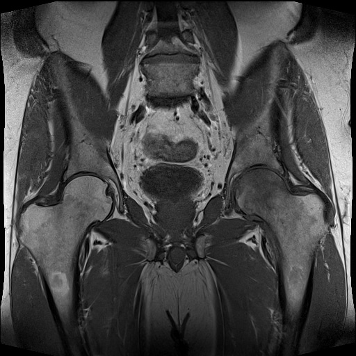
Figure 1. MRI scan (T1 images) of a normal hip (left) and bone changes in the femoral head in TIOsteoporosis (right).
TIO generally resolves over a period of months (3 – 12) without any specific treatment. When the hip is painful, simple painkillers may be helpful and the use of crutches can help both pain and protect the bone from damage whilst it is in a weakened state. Simple exercise to maintain flexibility and strength of the hip girdle muscle groups (gluteals, flexors and core) can be undertaken once the acute pain subsides. Aquatic exercises can be very helpful in this regard.
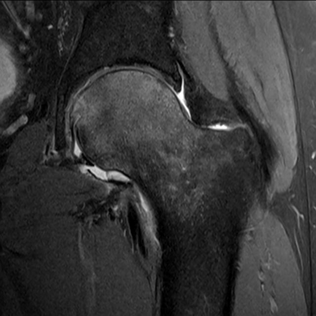
Figure 2. MRI scan (T2 images) of TIO of the Femoral Head. Bone oedema changes secondary to osteoporosis appear light in colour, whereas normal bone is dark in these images.
Source: https://www.parkclinic.com.au/transient-idiopathic-osteoporosis-of-the-hip/