Arthritis is a condition that can affect any joint in the body.
A joint forms between the ends of 2 bones and is the structure that allows movement – ie The Knee joint is a complex joint where the femur bone (thigh) interfaces with the tibia (shin) bone and the patella (kneecap) articulates with a groove on the front surface of the femur.
The joint surface is a specialized layer of gristle like tissue 2-3 mm thick that sits on the end of the bone. The scientific name for this layer is articular cartilage. This tissue acts like a shock absorber and reduces load on the bone beneath. It also has very low friction characteristics which allow the surfaces to glide easily over each other.
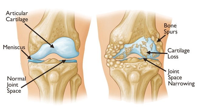
Arthritis is where there is loss (partial or complete) of this joint surface layer. This loss is usually progressive and ultimately leads to a situation where the underlying bone is exposed or uncovered resulting in “bone on bone” contact – this is the situation in an advanced or end stage arthritis. A good analogy for arthritis is a car tyre where the tread has worn down.
Figure 1. Normal Knee (left) and Arthritic Knee (right) with articular cartilage damage.
The main symptom associated with arthritis is pain. The exact cause of pain in arthritis is uncertain and probably multifactorial. Bone is a very sensitive tissue with lots of nerve endings – a broken bone is usually very painful. When the bone is not properly protected by a damaged joint surface, the bone is subject to more load and stress, which can cause pain. The wear debris created by joint surface damage floats around the joint and irritates and inflames the lining tissue of the joint (synovial lining) which also can be a contributory cause for pain.
Figure 2. Arthroscopic views of the Knee. Normal articular cartilage joint surfaces and meniscus (left). Mild – moderate articular cartilage damage (middle). Severe articular cartilage loss with bone exposed on joint surfaces (right).
Read more about osteoarthritis of the knee.

